
Panoramic dental imaging that shows the map beneath the smile.
OPG and CBCT scans help us see roots, bone, wisdom teeth, sinuses, nerves, infections, and implant sites before treatment decisions are made.
First, choose the question
The right scan depends on the decision being made.
Broad screening, implant planning, wisdom tooth removal, and single-tooth diagnosis do not all need the same image.
Scan type
OPG or CBCT
Useful for
Hidden anatomy
Visit feel
Quick standing scan
Afterward
No recovery time

What imaging does
A scan helps explain what the eye cannot see.
Teeth are only part of the story. Dental imaging can show root shape, bone support, infection patterns, impacted teeth, sinus position, and anatomy that affects treatment safety.
Width
Full jaw view
Depth
3D when needed
Plan
Treatment route
Scan types
Different images answer different dental questions.
The safest and clearest approach is to choose the image that gives enough information for the decision, without scanning more than needed.
Panoramic OPG
A wide 2D image that captures the upper jaw, lower jaw, teeth, roots, wisdom teeth, jaw joints, and parts of the sinuses in one view.
Often used for
General screening, wisdom teeth, missing teeth, broad bone changes
CBCT 3D scan
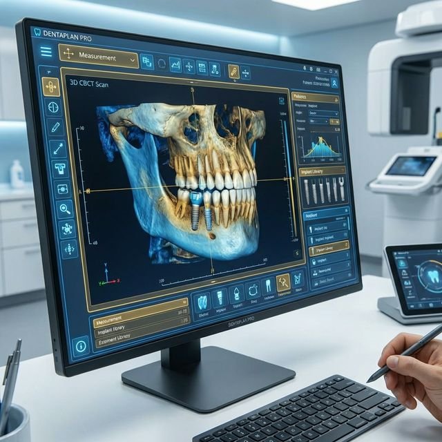
A three-dimensional scan used when depth, bone width, nerve position, sinus anatomy, or complex root shape needs more detail.
Often used for
Implants, surgical planning, impacted teeth, complex root or bone questions
Small dental X-ray
A focused image may be enough when the question is limited to one tooth, one filling, one root, or a small area of decay.
Often used for
Single-tooth diagnosis, bitewing decay checks, root canal follow-up
When imaging helps
A scan is useful when anatomy changes the plan.
We recommend panoramic or 3D imaging when it can reveal information that affects diagnosis, risk, timing, or treatment design.
Wisdom tooth planning
Imaging shows tooth angle, root shape, available space, and how close the tooth may be to important structures.
Implant assessment
CBCT helps evaluate bone height, width, density, sinus position, and nerve location before implant planning.
Unexplained pain or infection
A scan can reveal infection patterns, root concerns, bone changes, or hidden problems that are difficult to see clinically.
Orthodontic or aligner planning
A broad view helps identify impacted teeth, missing teeth, jaw relationships, and root positions before movement starts.
Before complex extractions
Root curves, tooth position, bone shape, and nearby anatomy can affect how treatment should be planned.
Reviewing old dental work
Imaging can help assess crowns, bridges, root canal treatment, implants, bone levels, and changes around older restorations.
Anatomy map
What we look for on the image.
The scan is reviewed in relation to your symptoms, exam findings, and planned treatment. The important finding is the one that changes what should happen next.
Impacted or unerupted teeth
Root shape and root direction
Bone level around teeth
Infection near root tips
Jaw cysts or unusual changes
Sinus position and sinus floor
Nerve canal location
Implant planning measurements
Your scan visit
A short appointment, planned around one clear question.
Define the question
We first decide what the image needs to answer, because different dental questions need different image types.
Position carefully
Your head and bite are positioned so the scan captures the right anatomy clearly and avoids unnecessary repeat images.
Capture quickly
The machine moves around your head while you stay still. The scan itself usually takes only seconds once positioned.
Review findings
Your dentist reviews the image, explains relevant findings, and connects them to diagnosis or treatment planning.
What scans cannot do alone
The image is evidence, not the whole diagnosis.
A scan becomes useful when it is matched with your symptoms, examination, dental history, and treatment goal.
A scan does not replace the exam
Symptoms, clinical tests, gum checks, bite checks, and medical history still matter. Imaging is one part of diagnosis.
Not every problem needs a large scan
A small dental X-ray may be better for a single cavity or one tooth. We choose the smallest useful image for the decision.
Some findings need comparison
Bone changes, healing, or old root canal areas may need earlier records or follow-up images to understand whether they are changing.
Safety and records
Imaging should be purposeful, explained, and stored properly.
Justified imaging
We recommend imaging when it can change diagnosis, treatment planning, referral, or monitoring.
Smallest useful view
For CBCT, the field of view is selected around the area that needs detail rather than scanning more than necessary.
Pregnancy check
Tell us before imaging if you are pregnant or may be pregnant so the dentist can decide the safest timing and approach.
Digital records
Digital images can support referrals, comparison, surgical planning, and future treatment decisions.
Planning examples
How imaging can change the treatment route.
Implant route
Bone and nerve location can decide implant size, position, need for grafting, or whether another replacement option is safer.
Wisdom tooth route
Root position and proximity to the nerve help decide whether monitoring, simple removal, or surgical extraction is appropriate.
Root canal route
Complex roots, infection size, missed canals, or previous treatment can be evaluated before deciding whether retreatment is sensible.
Orthodontic route
Impacted teeth, missing teeth, bone boundaries, and root positions affect what movement is realistic.
What affects imaging cost?
Cost depends on the type of scan, the area captured, whether detailed measurements are required, and whether the image supports a larger treatment plan.
The value is not the picture. It is the decision it supports.
A useful scan can help avoid surprises, reduce surgical risk, plan implants more carefully, explain pain, or confirm whether a simpler image is enough.
Book the Right ScanQuestions patients ask
Panoramic imaging FAQs
Book imaging
Start with the scan that answers the right question.
Tell us why imaging was recommended, what treatment you are planning, or what symptom needs investigation. We will guide the scan choice around that decision.